

We employ the latest technology to craft state of the art microscopy devices. By focusing on the researchers’ needs we are able to address their demands and tailor our microscopes to the latest standards in the industry.
Explore
the Unseen
Telight is specialist in the field of instruments employing light optics, in particular, providing innovative types and solutions for light microscopy. Live cell imaging is the heart of our company, we aim provide tools which will help researchers to advance in their exploratory work.
 January 24, 2024
 Imaging services for Discovery and Translational Research 2024
DATE: Apr 17-19 Join us together with Peter at Torino for the first NODE meeting, which we are attending this year; it is organized by the European BioImaging Industry Board.
 January 31, 2024
 Cell migration conference 2024
DATE: APR 15-18 We are going to join a unique event focused on different aspects of cell migration for the first time.
 January 31, 2024
 ELMI 2024 | European Light Microscopy Initiative
DATE: 4 - 7 JUN We will be going to the renowned conference European Light Microscopy Initiative 2024. In the last year, we have improved our devices and software; we are keen to share our advancements with the light microscopy community!
Telight Q-Phase
Quantitative Phase ImagingQ-Phase is a patented holographic microscope with high detection sensitivity.
Q-Phase is an ideal solution for experts who desire precise automated segmentation of individual cells for subsequent data analysis. Q-Phase quickly transforms cell features and dynamics into numerical data suitable for comparisons, correlations, and more detailed statistics.
Read more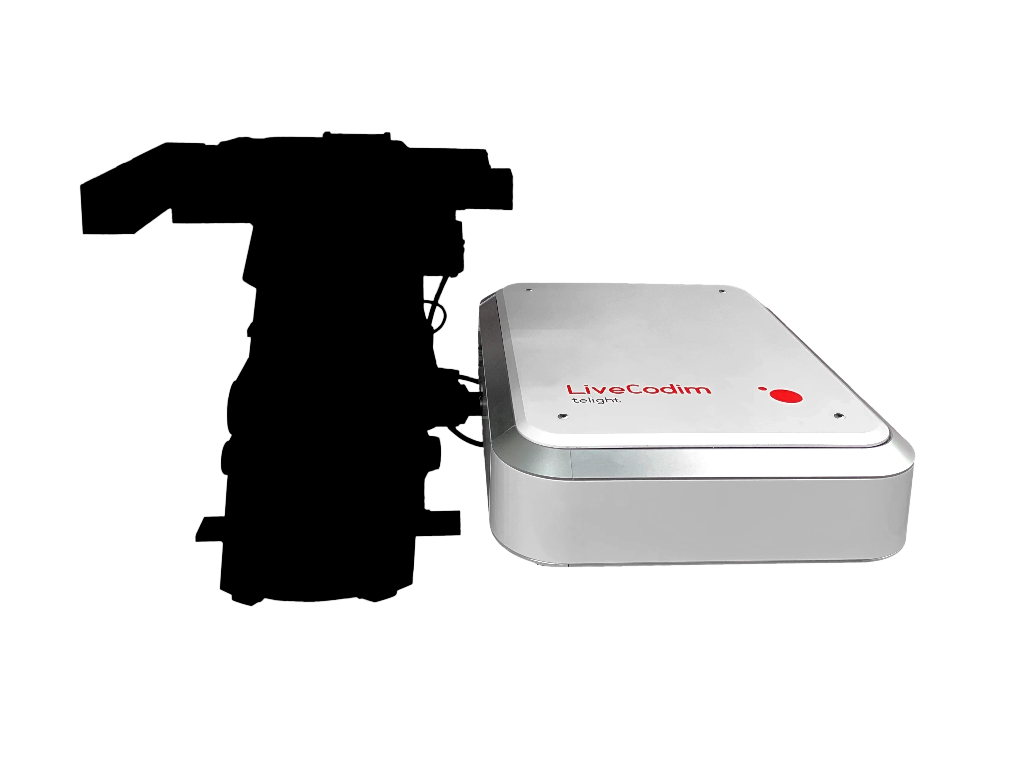
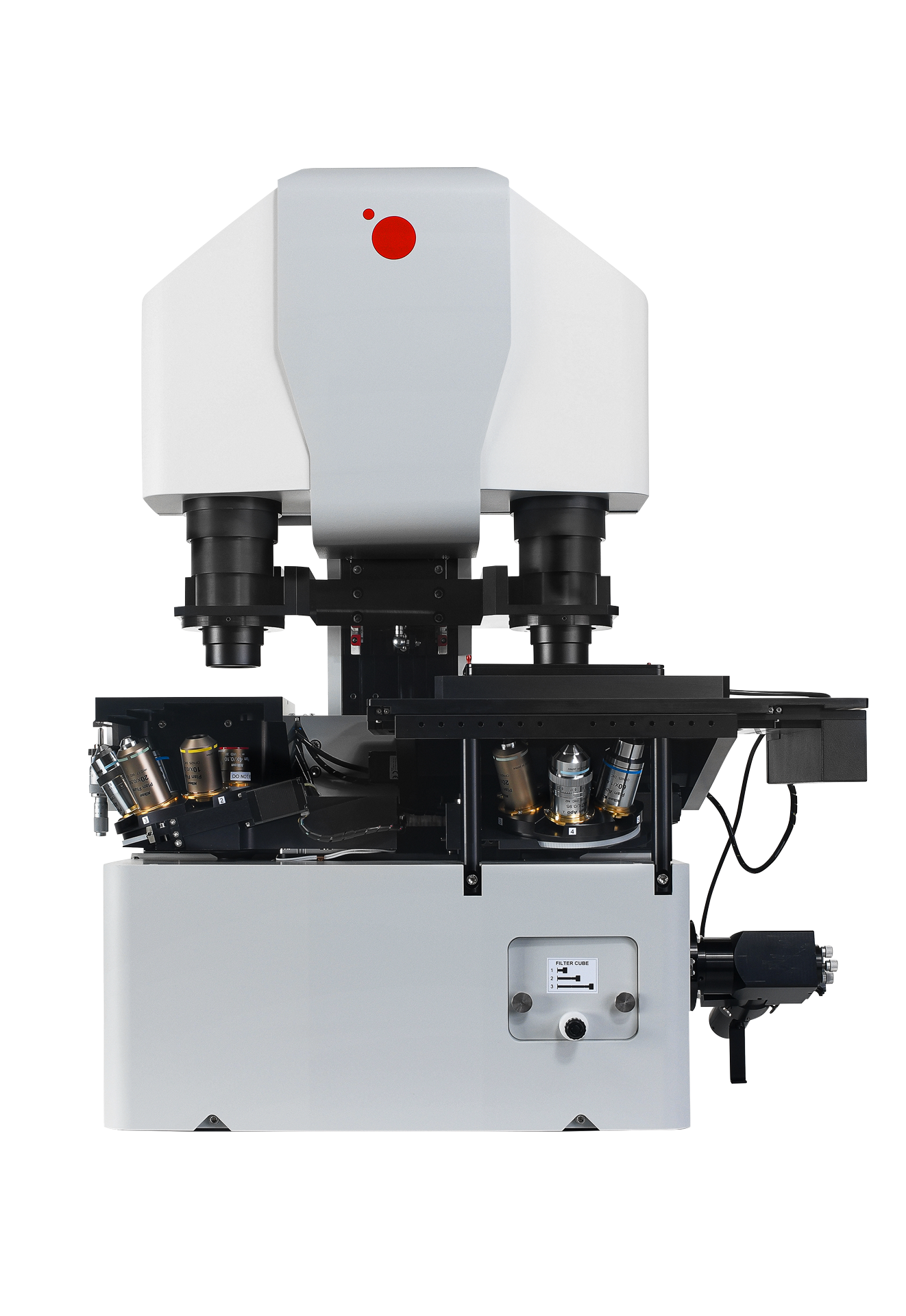
Telight LiveCodim
From conventional to super-resolution microscopy
LiveCodim is a universal, super-resolution imaging platform designed to interface with any standard fluorescence microscope. It is the solution for live-cell imaging with high resolution and low phototoxicity.
Read more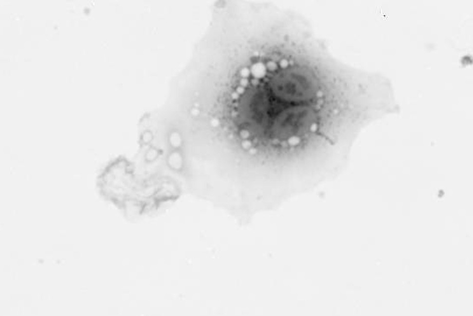
Testimonials

Our research group devised the holographic principle of the Q-Phase Multimodal microscope and participated in the development and testing. Currently, we use it in cell biology and nanomaterials research. We exploit the extraordinary properties of incoherent quantitative phase imaging particularly in cancer research by looking for a new kind of biomarkers relevant to personalized tumor treatment and also useful in a search for migrastatics. A very important application area is the investigation of optical properties of meta-surfaces. Moreover, with Q-Phase microscope we can test new imaging modes that incoherent holography offers such as imaging objects in optically scattering environments, imaging 3D objects, and new methods of super-resolution.
Radim Chmelík
Experimental Biophotonics Research Group Leader, CEITEC BUT

The quantitative phase imaging is the feature I appreciate the most. It offers high quality images and quantitative data, which cannot be easily obtained elsewhere. I also appreciate the possibility to combine phase imaging with the fluorescence microscopy. I like effective technical support, too.
Tomáš Groušl
Independent researcher (Laboratory of Cell Signalling)

The biggest advantages of using Q-PHASE include relatively robust data recruitment, automation of the acquisition process, combination of fluorescence and QPI, and high resolution.
Jan Balvan
Assistant professor, Department of Physiology

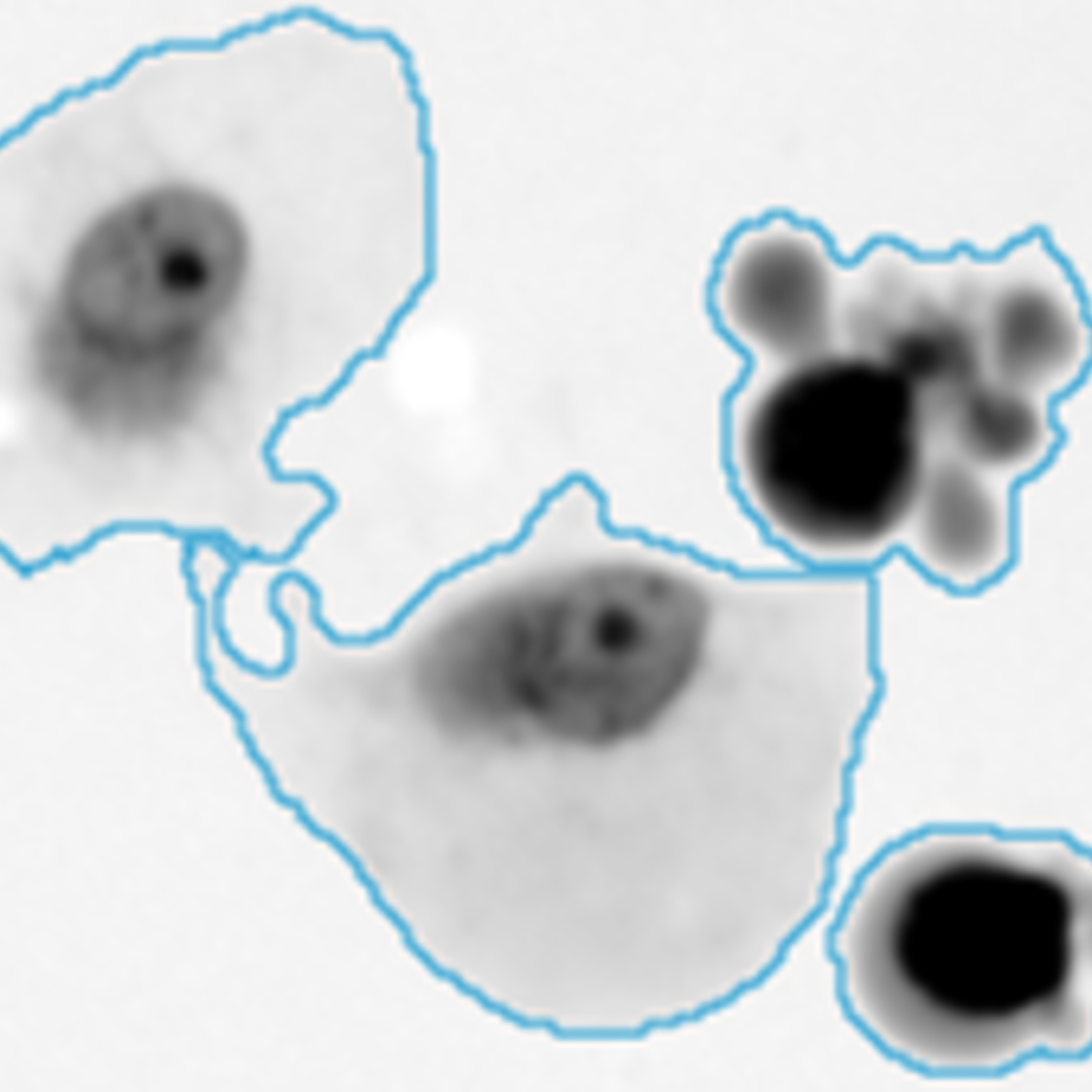
Using the method of dynamic phase differences provided by Q-Phase, our laboratory in collaboration with Prof. Chmelík’s laboratory described the dynamics of differences in cell mass distribution in mesenchymal and amoeboid tumor cells migrating in a 3D environment. We have also shown, using this method, that some features are common to both modes of invasion. We found that blebbing is enhanced in amoeboid fibrosarcoma cells as they pass through small pores, and that blebbing is occasionally present in mesenchymal-invading cells around nuclei that are compressed by surrounding collagen fibers. We further demonstrated that both the leading processes and leading panicles of invading fibrosarcoma cells are defined by higher cell mass density. In addition, we directly documented the fusion of collagen fibers by the protrusions of mesenchymal fibrosarcoma cells. Thus, the type of non-invasive microscopy presented here offers new insights into cellular events during 3D invasion and contributes significantly to our understanding of this crucial step in the metastatic process.
Doc. RNDr. Jan Brábek, Ph.D.
Leader of the Molecular and Cellular Mechanisms of Invasiveness of Tumour Cells group at the BIOCEV